
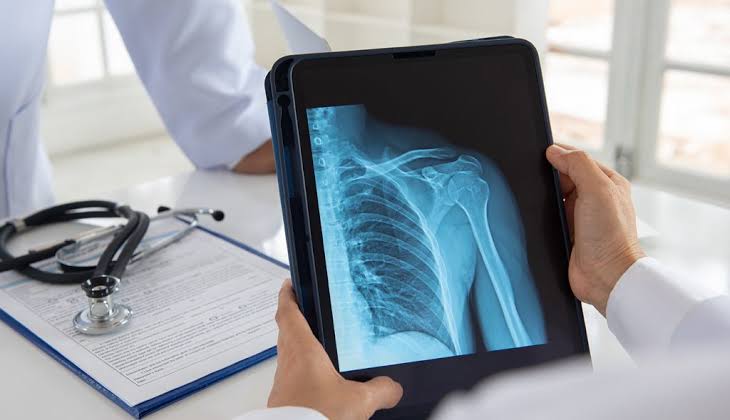
How Digital Radiography Is Improving Speed, Quality and Patient Care!.
Medical imaging has changed significantly with the shift from traditional film-based X-rays to digital technologies. A digital radiography system uses digital detectors to capture X-ray information and convert it into images that can be viewed on a computer. Unlike computed radiography, DR captures the X-ray signal electronically and makes the image available almost immediately. This can support faster workflows and reduce the need for physical film handling.
How DR Imaging Works
DR imaging starts when X-rays pass through the patient’s body and reach a digital detector. The detector converts the received information into digital data, which is processed by imaging software and displayed for review. Modern systems can include the X-ray source, detector, image processing software and acquisition workstation as part of an integrated workflow.
This process makes DR imaging useful in hospitals, diagnostic centres and other healthcare environments where quick access to radiographic images is important.
Improving Radiology Workflow
One of the major benefits of a digital radiography system is workflow efficiency. Images can become available shortly after exposure, allowing technologists to check them without waiting for film development or cassette-based processing. Digital images can also be transferred, stored and accessed through connected medical imaging systems.
For busy departments, efficient DR imaging can help reduce unnecessary delays between image acquisition and interpretation. Better workflow does not only mean speed. It also requires proper training, quality control and consistent imaging protocols.
Image Quality and Dose Management
Image quality is an important part of digital radiography. Detector performance, exposure settings, positioning and image processing can all affect the final image. The American College of Radiology recommends using digital radiography practices that provide appropriate image quality while maintaining suitable radiation exposure.
Radiation dose should always be optimized according to the examination, patient size and clinical requirement. Regular monitoring can help imaging teams identify unusual exposure patterns and improve protocols when necessary.
Choosing Modern DR Radiology Solutions
Healthcare facilities looking at DR radiology solutions should consider more than image acquisition alone. Detector reliability, image processing, workflow integration, usability, service support and quality assurance are important factors. The right solution should fit the department’s examination volume and clinical requirements.
Well-designed DR radiology solutions can also support consistent imaging across different examinations. Features such as exposure monitoring and standardized workflows can contribute to quality improvement when properly implemented.
Supporting the Future of Radiology
As healthcare becomes increasingly digital, DR imaging continues to play an important role in routine X-ray services. A digital radiography system can support faster image access, digital storage and smoother communication between imaging professionals. At the same time, DR radiology solutions need to be selected and operated with attention to patient safety, image quality and long-term workflow requirements.
Conclusion
Modern radiography is not simply about producing an X-ray image faster. It is about creating a dependable imaging workflow that brings together technology, trained professionals, quality assurance and patient-focused care. A strong imaging technology provider can support healthcare facilities with reliable digital platforms, advanced image processing and practical workflow solutions designed around everyday radiology requirements.